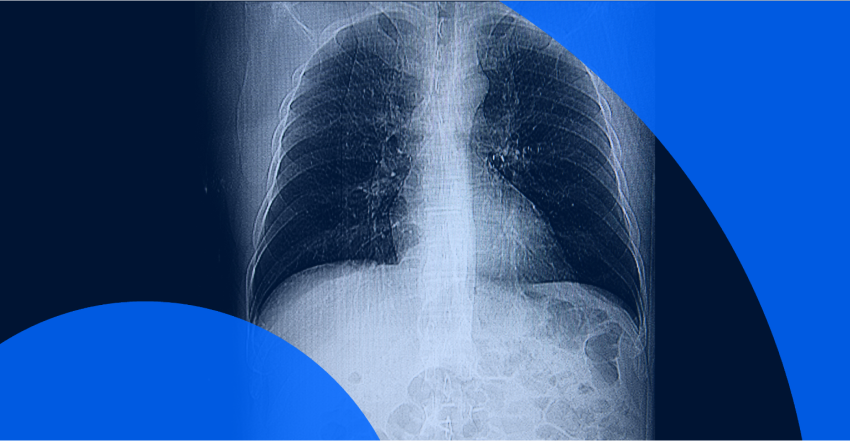
A CT scan creates detailed images of the internal structures and workings of your body using X-rays. A doctor might refer you for a CT scan to diagnose, identify or monitor the treatment of illnesses or injuries.
In Canada and the U.S., you have the right to access your health information contained in medical or health records. Having access to your medical records, including CT scan results, allows you to advocate for your own health and treatment, while also relieving ‘scanxiety’, the stress of waiting for medical results. A 2023 Leger study revealed more than half of patients experience ‘scanxiety’ when waiting on medical results.
With PocketHealth, you can get early access to your medical images and reports, often as soon as they’re released by the hospital imaging department or clinic.
But even when you have access to your health records, that doesn’t mean they’re simple to read or understand. Medical reports are still full of complex terminology. This article will clarify some of the medical terms and abbreviations you might encounter in your CT scan report so you’ll know where you stand and can be more prepared for conversations with your healthcare team.
CT is short for computed tomography. Also known as a CAT scan, a CT scan uses a donut-shaped array to take multiple X-rays from many different angles, creating a 2D cross-sectional “slice” at one location within the body. These slices are then collected at multiple contiguous locations and combined digitally into a detailed 3D image.
A radiologist will interpret these images (sometimes hundreds or thousands of images in one exam) and create a detailed report. Most commonly, your referring physician will receive the radiology report and discuss the results with you at a follow-up appointment.
A CT scan provides much more detail than a single X-ray and can be used to locate clots and tumors, identify bone or muscle issues, examine organs, diagnose specific illnesses and monitor treatment.
A CT scan report may seem complicated, but once you understand the main principles it will be much more comprehensible to read. Below we’ll explain planes, density, the Hounsfield scale and other important components you’ll find helpful in interpreting your CT scan report.
The term “plane” is used to describe the viewing angle of a patient in a CT scan image. Images are obtained in 1 plane and then reconstructed along 3 different planes to provide the radiologist with a 360° view of your body’s internal structures and organs:
Some CT scans require a contrast agent, a liquid composed of iodine that works by making structures with contrast appear more white (see “density” below). In a CT scan with contrast, an abnormality may become more obvious, either because it appears denser with contrast (enhancing/hyper-enhancing) or because the surrounding tissue becomes brighter, outlining the abnormality (hypodense).
Contrast for a CT scan is most commonly administered in 3 ways:
A CT scan with contrast might make use of the multiphase imaging technique, which takes images at specific points in time, for instance as the contrast reaches specific blood vessels or moves through specific organs.
CT scan images show up in shades of gray, from white to black. These shades measure the density (or lucency) of each of your internal structures compared to the next. As X-rays pass through a person they are absorbed in varying degrees (depending on the material of the bones, tissues or organs they encounter) before being captured digitally.
A way of quantifying the absorption of X-ray beams as seen on CT scans was first described by Sir Godfrey Hounsfield. Tissues that absorb X-rays are termed dense and appear white; conversely, tissues that do not absorb X-rays are termed lucent and appear black. Here’s a breakdown:
The Hounsfield scale measures tissue density and assigns a common scale to express how different tissues in your body absorb the radiation from the CT scan. This process of absorption is called attenuation.
Hounsfield units (HU) are standard numbers on the scale that express the radiodensity of different parts of the body, relative to water (assigned as zero). Your CT scan report may contain HU scale numbers when certain masses or cysts are reported.
The lower the number on the Hounsfield scale, the less dense the tissue is. The higher the number, the more dense the tissue is. For example:
Based on the radiologist’s review of the CT scan images, they’ll determine the presence of any abnormalities (often termed “findings” or “observations” in radiology lingo). Some descriptors of findings you may see in your radiology report to communicate these abnormalities include:
Depending on their source and composition, any of these findings can be benign (non-cancerous) or malignant (cancerous). Some imaging abnormalities can be diagnosed definitively with a CT scan, while other abnormalities would need to be proven using a tissue biopsy.
When it comes to medical terminology the term “unremarkable” is actually a good thing! It means your CT scan has revealed no significant abnormal findings.